
Position:Home >
Lower Extremity Venous Ulcer (VLU) Model
Lower Extremity Venous Ulcer (VLU) Model
Background
Lower extremity venous ulcers are a common and frequently encountered condition in surgical practice, representing one of the primary clinical manifestations of advanced chronic venous insufficiency in the legs. In traditional Chinese medicine, they are termed “leg sores” or “chronic leg ulcers.” This condition can lead to chronic pain, disability, and reduced quality of life. Common sites of occurrence include the anterior medial aspect of the mid-to-lower calf (the boot area), followed by the
Materials and methods
Healthy SPF-grade SD rats (male, approximately 7 weeks old, weighing 240–290 g)
Model creation method: Anesthesia, skin preparation, femoral vein thrombosis induction, establishment of rat wound ulcer model.
Evaluation Criteria: Thrombus formation in the mold; Doppler flow measurement; Changes in lower limb ulcers; Hematoxylin and eosin staining of ulcer areas
Model creation method: Anesthesia, skin preparation, femoral vein thrombosis induction, establishment of rat wound ulcer model.
Evaluation Criteria: Thrombus formation in the mold; Doppler flow measurement; Changes in lower limb ulcers; Hematoxylin and eosin staining of ulcer areas
Test and verify
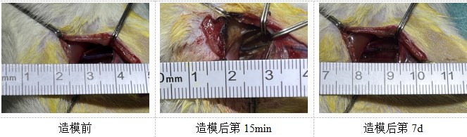
造模血栓形成
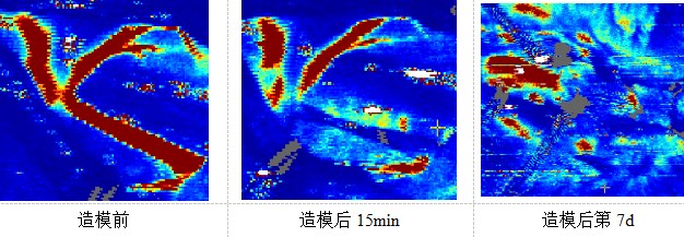
多普勒血流测定
溃疡形成情况
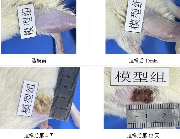
在烫伤处理15min后,模型组皮肤苍白,烫伤处理后第4天开始出现结痂,后期结痂情况逐渐加重,符合造模趋势。
溃疡区域HE染色
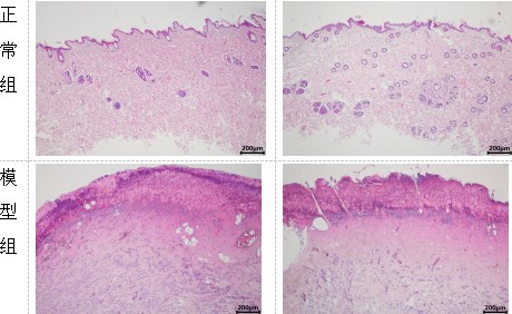
正常组组织结构正常,表皮层与真皮层结构清晰,未见明显病理变化;模型组样本结构严重受损,表皮层消失,同时伴随大量炎性细胞浸润与纤维结缔组织增生,病变严重,模型成功。

